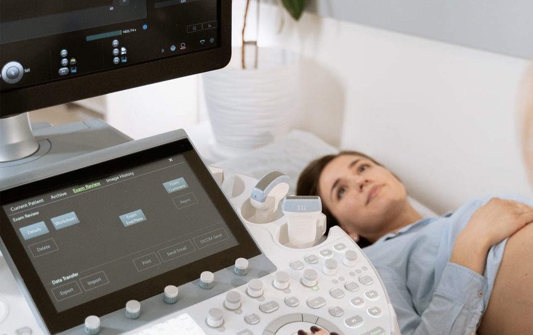
Hearing a baby’s heartbeat for the first time is an important milestone for new parents. And almost every parent wonders when they can hear their baby’s heartbeat.
Every woman and every pregnancy develops differently. Similarly, every baby and their developmental progress is different.
That being said, according to experts, you can generally start to see visible cardiac activity on an ultrasound around 5.5 to 6 weeks.
In this article, you will read everything you need to know about your baby’s heartbeat.
When does a baby have a heartbeat?
According to the American College of Obstetricians and Gynecologists (ACOG), a baby’s cardiac tissue, which will then form the heart, starts to develop within the first 8 weeks of pregnancy.
Cardiac tissue starts to pulse at around 5.5-6 weeks of pregnancy, registering as a heartbeat on the ultrasound, though the heart has not developed yet.
The heart of a fetus is fully developed by the 10th week of pregnancy.
Table below displays the development of the heart in weeks 5-10 of pregnancy as per medicalnewstoday experts.
|
Week of pregnancy |
Level of heart development |
|
Week 5 |
The developing heart is made up of two tubes that are fused in the middle, creating a trunk with four tubes branching off. Cardiac tissue begins to contract, and it may be possible to detect it using a vaginal ultrasound. |
|
Week 6 |
The heart of the embryo changes dramatically — the basic heart tube loops, forming an “S” shape. |
|
Week 7 |
The pumping chambers, or ventricles, and receiving chambers, or atria, of the heart begin to separate and develop. |
|
Week 8 |
The valves between the atria and ventricles of the heart form. |
|
Weeks 9 and 10 |
The aorta and pulmonary vein form. By week 10, the fetal heart has developed fully. |
When can I hear my baby’s heartbeat?
You may hear cardiac activity for the first time from week 5.5-6 of pregnancy or later, if you have an ultrasound at one of your early prenatal appointments. But always keep in mind that the timing of when it can be detected can vary a bit.
What if you can’t hear the heartbeat yet and it’s already week 6? Doctors recommend not to worry. At your next appointment, your doctor will check to make sure everything is okay.
Later on, at your 20 week ultrasound which is also called the level 2 ultrasound, you’ll hear and see your baby’s heartbeat.
A level 2 ultrasound focuses on fetal anatomy to ensure that everything is growing and developing as it should.
At level 2, ultrasound images are much clearer and more detailed than the ultrasound you might have gotten in your first trimester.
What is a normal fetal heart rate during pregnancy?
Experts measure fetal heart rate by the number of fetal heartbeats per minute (BPM) during pregnancy.
This measurement helps to determine the well-being of the fetus during prenatal visits or labor.
For most of your pregnancy, the fetal heart rate will be between 110 and 160 BPM.
Does fetal heart rate change throughout pregnancy?
The average fetal heart rate varies depending on the stage of your pregnancy.
- Weeks 5 to 7: A baby’s heart starts to develop around the 5. week of pregnancy. In this early stage, the heart rate is slower, averaging between 90 and 110 BPM.
- Week 8 to 12: The heart rate speeds up. The average is 140 to 170 BPM by week 9. By week 12, the rate slows down a bit.
- Week 13 to 26: Throughout most of the pregnancy, the average is 110 to 160 BPM. Week 27 to 40: During the last trimester, the fetal heart rate continues to average at 110 to 160 BPM. However, it drops slightly in the last 10 weeks. In general, it moves toward the lower end of this range the closer you get to your due date.
How does fetal heart rate change throughout the day?
Fetal heart rate can vary throughout the day and night by as much as 5 to 25 beats per minute (BPM). This change is due to the fetus’ activity level.
- The heart rate increases while the fetus is moving around and decreases while the fetus is asleep. These changes are similar to what adults experience while exercising or at rest.
- Weeks 10 to 12 are typically when the fetus’ heartbeat can be heard for the first time during a prenatal visit.
How is baby’s heartbeat monitored?
There are numerous ways to monitor a baby’s heartbeat.
- Most doctors use transvaginal ultrasounds in the early period. These wand-like instruments do an internal scan of your organs, including your uterus—or womb.
- As the pregnancy progresses, doctors tend to use fetal Dopplers. These handheld devices can detect your baby’s heartbeat as early as 8 weeks. But variables like whether you carry any amount of weight around your mid-section and the position of your uterus might make it difficult to monitor at this early gestational age. Most fetal heart tones can be heard by 10-12 weeks.
- Regular ultrasounds may also be used to monitor a baby’s heartbeat.
How do your baby’s heart and circulatory system develop?
Fetal heart development begins early on in pregnancy.
Below you will find a rough timeline of heart and circulatory system development.
First trimester development
By week 4, a distinct cluster of cells form inside your embryo. These soon develop into your baby’s heart and circulatory (blood) system.
At week 5, the preliminary structures that will become your baby’s heart begin spontaneously pulsing.
In these early stages, it resembles a tube that twists and divides to eventually form the heart and valves (which open and close to release blood from the heart to the body).
Precursor blood vessels also begin to form in the embryo during the first few weeks.
Second trimester development
Around week 17, the fetal brain begins to regulate the heartbeat in preparation for life outside the womb.
Capillaries also form at an exponential rate during the second trimester. These teeny-tiny blood vessels deliver oxygenated blood to the tissues in your baby’s body and then recycle deoxygenated blood back into the circulatory system.
Between 17 and 20 weeks, the heart chambers develop enough to appear more clearly on an ultrasound.
During your second trimester scan, your doctor will check the structure of your baby’s heart and look for any congenital heart defects.
If needed, your doctor may recommend a fetal echocardiogram between 18 and 24 weeks of pregnancy.
Third trimester development
A baby’s circulatory system continues to grow slowly and steadily during the last trimester.
How does a fetal heart work?
While the fetal circulatory system develops rapidly throughout pregnancy, it actually works quite differently in utero than it does once your baby is born.
Babies don’t breathe in utero and their lungs don’t actually function before birth.
Until then, your baby’s developing circulatory system relies on the umbilical cord for a steady supply of oxygen and nutrient-rich blood.
Umbilical arteries and veins transport what your baby needs from your body to theirs, then carry unoxygenated blood and waste products back to you for removal.
References: whattoexpect.com, healthline.com, acog.org, verywellhealth.com, parents.com, hopkinsallchildrens.org, thebump.com


